利用细胞系进行纳米材料体外生物效应的评价是纳米材料生物效应研究的一种重要手段。然而越来越多的研究认为,一些细胞系的过度传代培养将影响其细胞特性。基于此,我们将融合基因BCR-ABL细胞转染永生化小鼠前B细胞Ba/F3,使其不再依赖于IF-3的生长。然后进行连续传代培养研究其稳定性以及其对纳米银的细胞反应性。结果显示,短期传代不会影响Ba/F3-BCR-ABL细胞的稳定性,纳米银诱导不同代数细胞效应没有显著差异。因此,使用这样培养的细胞进行细胞效应评价时可以提供一致结果。
GUO DaWei, ZHANG XiuYan, HUANG ZhiHai, ZHOU XueFeng, ZHU LingYing, ZHAO Yun, GU Ning,Comparison of cellular responses across multiple passage numbers in Ba/F3-BCR-ABL cells induced by silver nanoparticles, SCIENCE CHINA (Life Sciences) 2012, 55(10), 898-905.

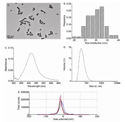
Figure 1Characterization of silver nanoparticles. A and B, The morphology and size distribution of the silvernanoparticles were characterized by transmission electron microscopy (TEM). The mean diameter was 29.7 nm. C, TheAgNPs were characterized by the UV-Vis absorption spectrum, and the maximum absorption was 417 nm. D and E,Particles size (diameter) and surface charge in an aqueous solution were measured by dynamic light scattering (DLS) andwere (71.83±0.74) nm and (23.0±1.01) mV, respectively.

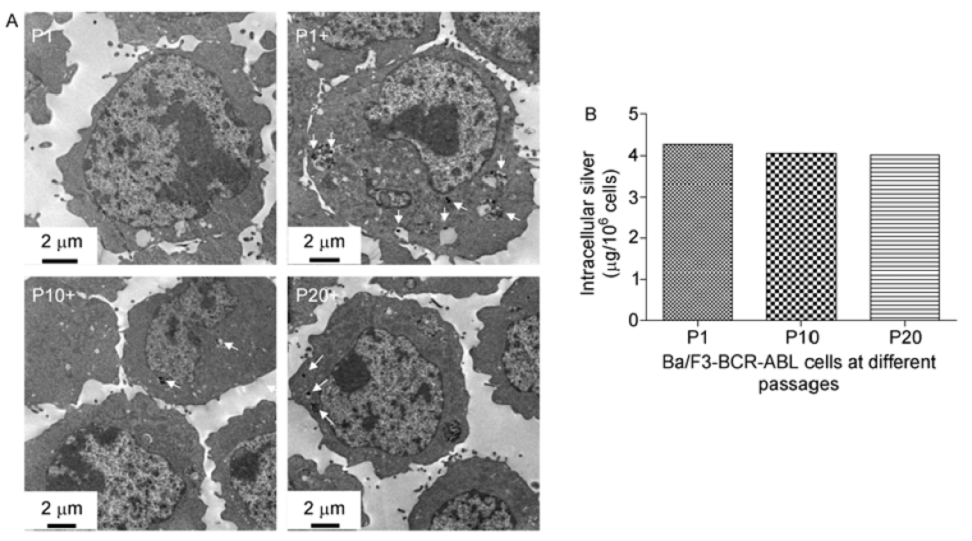
Figure 4Uptake and localization of AgNPs in Ba/F3-BCR-ABL cells at multiple passage numbers. A, TEM images ofultrathin sections of Ba/F3-BCR-ABL cells treated with AgNPs (12μg mL1) for 24 h showed that the particles wereinternalized and could be sequestered in vacuoles, which were visible in the cells as black, and the electron-dense spots areindicated by arrows. B, The cells treated with AgNPs (12μg mL1) for 24 h were digested in nitric acid, and the silver concentration was determined by ICP-MS. “+” denotes that cells were treated with AgNPs.
Figure 5 Effects of AgNP exposure on Ba/F3-BCR-ABL cell viability atmultiple passage numbers. After a 24-hexposure of AgNPs at multiple concentrations, cell viability was determined with a Cell Counting Kit-8 (CCK-8). The datawere a summary of three independent experiments and expressed as the means±SD. * denotes a significant reduction incell viability (P<0 .05) compared with the control.
Figure 6Effect of passage number and AgNP exposure on apoptosis of Ba/F3-BCR-ABL cells. A and B, Therepresentative images and summaries of apoptosis of the AgNP-treated (10μg mL1) and untreated Ba/F3-BCR-ABLcells at multiple passage numbers are presented. C and D, The representative images and summaries of apoptosis of the 1st passage Ba/F3-BCR-ABL cells induced by AgNP exposure at various concentrations are presented.
Figure 7Effect of passage number and AgNP exposure on the cell cycle of Ba/F3-BCR-ABL cells. A and B, Therepresentative images and summaries of the cell cycle of the AgNP-treated (10μg mL1) and untreated Ba/F3-BCR-ABLcells at multiple passage numbers are presented. C and D, The representative images and summaries of the cell cycle of the 1st passage Ba/F3-BCR-ABL cells induced by AgNP exposure at various concentrations are presented.