通过同步辐射源、薄闪烁体以及光学系统搭建的高分辨CT系统因为其分辨率可以达到亚微米甚至十纳米量级。介于这个优势,它可以方便用于组织细胞成像。这个系统中要求的闪烁体厚度通常只有几个微米甚至亚微米,这种厚度的闪烁体加工难度很大,同时,较薄的闪烁体厚度会大大的降低闪烁体的发光效率,增大样本受辐射剂量。怎样在高分辨的情况下降低闪烁体的厚度要求是一件非常有意义的研究工作。
本文提出的方法可以用于扩展闪烁体的系统景深,通过较厚的闪烁体代替较薄的闪烁体进行高分辨成像。文中我们首先从理论上推导出了由于景深原因导致的模糊图像和系统原始清晰图像之间的关系模型,继而通过凸集投射算法和全变差算法实现了模糊图像到清晰图像的重构。
实验中,我们模拟了一个闪烁体厚度要求是1微米的高分辨系统,并用20微米的闪烁体代替之。首先我们得到了由于不匹配厚度的闪烁体产生的模糊图像。通过我们的算法,在无噪声情况下,完全复原了原图像;在有噪声的情况下,恢复的效果良好,分辨率得到了很大的提高。但是低对比度图像的高质量恢复还是需要依赖于图像的信噪比。因此相应的去噪算法还值得今后更加深入的研究。
Guang Li, Shouhua Luo, Yuling Yan, Ning Gu*, A method of extending the depth of focus of thehigh-resolution X-ray imaging system employing optical lens and scintillator: a phantom study BioMedical Engineering, 2015, 14(Suppl 1):S15,doi:10.1186/1475-925X-14-S1-S15.
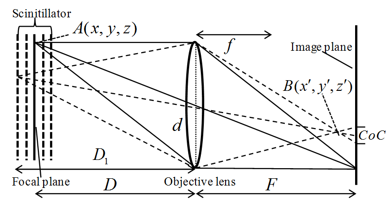
Imaging model of high-resolution X-ray imaging system in which the thickness of the employed scintillator is not matching with the objective lens.
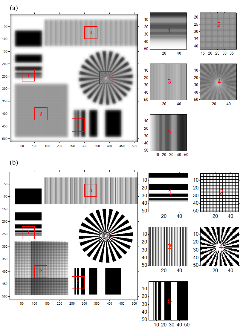
Comparison between the blur image and the result images from the proposed method: (a) simulated obscure image and its some parts with magnification, (b) recovery image and some parts of it with magnification.
